The eye is one of the structures in the body where time from symptom onset to treatment genuinely shapes the outcome. A corneal ulcer that is treated promptly heals cleanly; one that is left for two days can perforate. Sudden pressure changes in glaucoma produce irreversible damage in a matter of hours. A new cataract could just be aging, or it could be the first sign of diabetes. This is not meant to create panic about every squint and discharge, but to make the case for knowing which eye signs constitute a real emergency.
Cobb & Co. Veterinary Clinic in Elgin offers urgent care six days a week during open hours, and our transparent, client-centered approach extends to being direct about when something needs to be seen today versus tomorrow. Our urgent and emergency pet care covers ocular emergencies alongside everything else. If your pet’s eye looks wrong, book now right away.
At a Glance
- The eye runs on a tight clock; corneal ulcers that perforate, glaucoma that takes vision in 24 to 48 hours, and proptosis that loses the eye if not replaced quickly all turn on speed of treatment.
- Sudden vision loss, severe redness with pain, trauma, chemical exposure, a visible foreign object, or any pupil that does not respond to light all warrant same-day evaluation rather than waiting.
- Some conditions look like emergencies but are not (mild conjunctivitis, nuclear sclerosis, longstanding tear staining), and some look mild but are not (subtle squinting from a deep corneal ulcer, slight redness from early glaucoma).
- Daily looks at your pet’s eyes during normal handling, combined with prompt evaluation when anything seems different, protect long-term vision better than any single intervention.
How Do You Know When Your Pet’s Eye Is Uncomfortable?
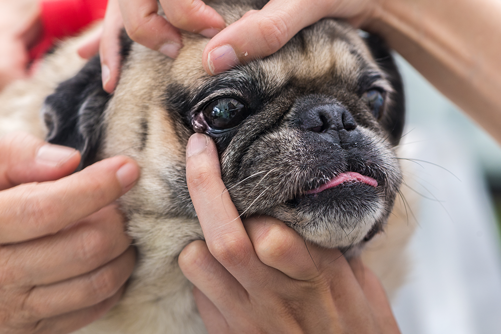
Pets are remarkably stoic about pain in general, and eye pain is no exception. The signs of eye pain are often subtle:
- Squinting or holding the eye partially closed
- Increased tearing or any colored discharge
- Redness of the white part of the eye or the inner eyelid
- Rubbing the face on furniture, carpets, or with paws
- Cloudiness, haziness, or any visible change in the surface of the eye
- Behavioral changes like withdrawal, decreased appetite, or reluctance to play
- Pawing at the affected eye
- A change in pupil size or shape
Any of these in your pet, if they were not there yesterday, warrant a phone call. Some are routine office visits; others are same-day emergencies. The point is not to wait and hope.
Which Eye Symptoms Need Same-Day Care?
When to Seek Immediate Veterinary Attention for Eye Injuries
Some eye emergencies cannot wait until a routine visit:
- Penetrating trauma (a stick, claw, or any object that has entered the eye)
- Chemical exposure (cleaning products, garden chemicals, anything alkaline or acidic)
- Sudden severe redness with apparent pain
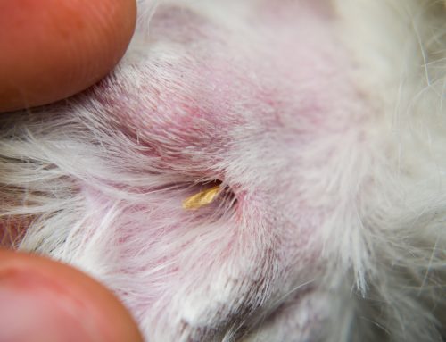
- A visible foreign object in or under the eyelid, including foxtails and other plant material
- Bleeding from the eye or surrounding tissue
- Eye swelling that is expanding or affecting vision
Practical guidance while heading our way:
- Do not try to remove a penetrating object yourself. It may be the only thing preventing more damage.
- For chemical exposure, gently flush the eye with copious clean water or saline for 15 to 20 minutes before transporting. This is one situation where on-the-spot first aid genuinely helps.
- Cover the eye loosely with a clean cloth if your pet will not tolerate flushing.
- Prevent self-trauma by putting an Elizabethan collar on if you have one
- Keep the carrier or vehicle calm and dim during transport. Bright lights are uncomfortable for an injured eye.
- Do not put any over-the-counter eye drops or ointments in the eye before being seen. Many human eye products contain ingredients that worsen specific eye conditions.
Which Eye Conditions Should Not Wait?
Eye conditions that are painful or that threaten vision should always be seen the same day. The conditions in this section are urgent because loss of vision (or in some cases, loss of the eye itself) is a real possibility.
Corneal Ulcers
Corneal ulcers are erosions of the surface layer of the cornea, the clear part of the eye. They are painful, and they are one of the more time-sensitive conditions we see. Causes include scratches from another cat, foreign bodies under the eyelid, dry eye that compromises the protective tear film, and underlying disease that affects healing.
Symptoms include intense squinting, copious tearing, a hazy or duller-looking eye surface, and obvious discomfort. Diagnosis is straightforward in our clinic with a fluorescein stain, which highlights any disrupted area of the cornea. Treatment depends on depth and complexity: superficial ulcers often heal with antibiotic drops and pain control over a week or two, while deeper ulcers require more aggressive intervention and sometimes surgical referral.
The progression from manageable ulcer to perforated eye can happen in days. Our sick pet visits accommodate same-day evaluation for any painful eye, which is the right way to handle a suspected ulcer.
Glaucoma
Glaucoma is increased pressure within the eye, and it is one of the most painful and time-sensitive conditions in veterinary ophthalmology. Vision loss begins within hours of pressure spikes, and by 48 hours, vision in the affected eye is often permanently gone.
Hallmark signs:
- A red, painful eye that often looks different from the other one
- Cloudiness or a hazy appearance to the cornea
- A dilated pupil that does not respond normally to light
- Squinting, severe pain, lethargy, decreased appetite
- The eye may appear larger than the other one in advanced cases
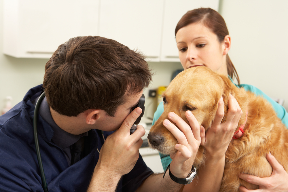
Glaucoma in either species is an emergency. Get to a clinic immediately. Our team measures intraocular pressure with a tonometer in-house, which gives us answers within minutes and lets us start emergency treatment promptly.
Lens Dislocation
Lens dislocation happens when the small fibers holding the lens in place weaken or break and the lens shifts forward or backward within the eye. The condition is most common in terriers (Jack Russells, Smooth Fox Terriers, Wirehaired Fox Terriers) and has a strong hereditary component in those breeds.
A forward-displaced lens can block the normal flow of fluid through the eye and trigger acute glaucoma, which means the urgency of lens dislocation often comes from what it causes rather than the dislocation itself. Signs include sudden onset of squinting, redness, a visibly displaced lens (sometimes you can see a crescent shape in the pupil), and the same painful presentation that suggests glaucoma. Treatment usually involves emergency pressure control and referral for surgical removal of the displaced lens in many cases.
Proptosis
Proptosis is the displacement of the eye out of its socket, almost always following trauma. Brachycephalic breeds (Pugs, Shih Tzus, Pekingese, Boston Terriers) are dramatically more susceptible because their shallow eye sockets and large eyes mean even moderate trauma can pop the eye out.
Proptosis is a true emergency. The longer the eye remains out of the socket, the worse the prognosis for vision and for saving the eye at all. Cover the eye with a clean, damp cloth (do not let it dry out) and head straight in. Do not try to push the eye back yourself. Surgical replacement and stabilization, performed quickly, gives the best chance of preserving function.
Hyphema
Hyphema is bleeding inside the front chamber of the eye, visible as a layer of red blood pooling against the iris or filling the eye entirely. The bleeding itself is not always the most dangerous part; the cause is. Hyphema can result from trauma, but it also signals serious underlying problems including high blood pressure, clotting disorders (immune-mediated thrombocytopenia, rodenticide toxicity), tumors inside the eye, or systemic disease.
Any visible blood in the eye warrants same-day evaluation. We work through the diagnostic process to identify the cause, treat the bleeding, and address whatever underlying issue is driving it. Blood pressure measurement and bloodwork are typically part of the initial workup.
Uveitis
Uveitis is inflammation of the middle layer of the eye (the iris and surrounding structures). It is painful, threatens vision through direct damage and secondary glaucoma, and is often a clue to systemic disease rather than an isolated eye problem.
Signs include redness, squinting, cloudy or hazy appearance to the eye, a constricted pupil (the opposite of the dilated pupil seen in glaucoma), and obvious discomfort. Causes are broad: infections (tick-borne diseases, fungal infections, feline leukemia virus, feline immunodeficiency virus, toxoplasmosis), cancer (lymphoma), immune-mediated disease, trauma, and lens-related disease. Workup includes a thorough eye exam plus systemic testing to find the cause, because treating the inflammation without addressing the source leads to recurrence.
Retrobulbar Abscess
A retrobulbar abscess is an infection in the space behind the eye, often related to dental disease, foreign material (grass awns are a classic culprit), or trauma. The eye itself usually looks normal at first glance, but the pet shows severe pain on opening the mouth, reluctance to eat, swelling around the eye or the side of the face, and sometimes a visibly protruding eye on the affected side.
Dental disease overall is one of the more underdiagnosed contributors to facial and periocular issues. Our dentistry services include comprehensive oral evaluation that catches these connections, especially in older pets where dental disease has had time to advance.
Sudden Vision Loss
Acute blindness (sudden vision loss with no obvious external eye changes) is one of the more alarming presentations we see. The eye may look completely normal, but your pet is suddenly bumping into furniture, hesitant on stairs, or unable to find toys they used to track easily. Causes range from reversible (hypertension causing retinal detachment in cats, which can resolve with prompt blood pressure control) to irreversible (some forms of retinal degeneration). Same-day evaluation matters because the causes that are reversible are reversible only if caught quickly.
SARDS (sudden acquired retinal degeneration syndrome) is a striking condition where dogs lose their vision over the course of days to weeks, often associated with subtle systemic symptoms like increased thirst, appetite changes, and weight gain. The diagnosis is made through specialized retinal evaluation, and unfortunately the vision loss is not reversible. Identifying the condition still matters, because it changes the conversation toward adaptation rather than continued treatment attempts.
Which Eye Conditions Need Care but Are Not Emergencies?
Some eye conditions should be seen promptly but do not necessarily require an immediate same-day visit. These can usually wait for the next available appointment, though prompt evaluation still matters.
Conjunctivitis
Conjunctivitis is inflammation of the membrane lining the eyelids and white of the eye, often called pink eye. It is common, and the cause matters because it shapes treatment.
- Allergic conjunctivitis: typically affects both eyes, produces clear or slightly mucoid discharge, and is often associated with seasonal allergy flares
- Bacterial conjunctivitis: tends to produce yellow or green discharge and may affect one eye initially
- Viral conjunctivitis: particularly feline herpesvirus in cats, can cause recurring flares and sometimes corneal involvement
- Irritant conjunctivitis: results from contact with chemicals, smoke, or foreign material
Treatment is matched to the cause. Bacterial conjunctivitis responds to antibiotic drops; viral cases may need antiviral medication; allergic cases often improve with addressing the underlying allergy.
Cherry Eye
Cherry eye is a prolapse of the gland of the third eyelid, which appears as a red, fleshy swelling at the inner corner of the eye. It is most common in young dogs of certain breeds (Bulldogs, Beagles, Cocker Spaniels, Cavaliers).
Beyond the cosmetic concern, the prolapsed gland produces a significant portion of the tear film. Leaving it untreated raises the risk of dry eye later in life, which is why surgical correction with gland preservation (rather than removal) is the modern standard. The prolapse also irritates the cornea and can lead to secondary problems if not addressed.
Cataracts
Diabetes is a major cause of cataracts in dogs, and they often progress quickly in diabetic patients. If your pet has other symptoms, like increased thirst and urination or weight loss in addition to a cataract, this bumps up to same-day evaluation. Differentiating a cataract from nuclear sclerosis is one of the most common confusions we see. Both produce a cloudy or bluish appearance in the eye, but they are very different things.
Cataracts vs. nuclear sclerosis differs in this way: nuclear sclerosis is a normal age-related change in the lens that produces a slight bluish haze in older pets but does not significantly affect vision. Cataracts are an actual lens opacity that disrupts vision, ranging from mild to complete. Nuclear sclerosis needs no treatment. Cataracts may require surgery to restore vision, particularly when both eyes are affected.
Dry Eye
Dry eye (keratoconjunctivitis sicca, or KCS) is a condition where tear production is inadequate to keep the eye lubricated. It is common in certain breeds (Cavaliers, Bulldogs, Pugs, Cocker Spaniels) and can develop secondary to autoimmune disease, certain medications, or prior eye surgery.
Hallmark signs include:
- Thick, ropy mucoid discharge
- Chronic redness and irritation
- Dull, lackluster eye surface
- Frequent corneal ulcers
- Cloudiness over time as chronic inflammation damages the cornea
Dry eye is diagnosed with a Schirmer tear test, which measures tear production. Treatment is lifelong: tear-stimulating medications (cyclosporine or tacrolimus) and artificial tears, with regular monitoring to adjust the plan over time. Our wellness and preventative services include rechecks for chronic conditions like dry eye where ongoing tracking matters.
For at-risk breeds, breed-specific screening and monitoring matters. We will talk through what is appropriate for your dog at routine wellness visits.
How Do You Care for Your Pet’s Eyes Routinely?
Most eye problems are easier to manage when caught early. A few habits make that more likely:
- Look at your pet’s eyes daily. A quick check during morning and evening interactions catches changes early.
- Wipe away normal tear staining with a damp cloth (not the same area used elsewhere on the body).
- Trim hair around the eyes in long-haired breeds to prevent constant irritation.
- Keep flat-faced breeds away from situations with high foreign body risk (running through tall grass, woodland brush).
- Schedule wellness exams regularly. Our team checks eyes at every visit, often catching subtle changes before you notice them.
Senior pets benefit from more frequent eye exams. The combination of age-related lens changes, retinal degeneration, increased glaucoma risk, and the higher likelihood of systemic disease that affects the eyes makes twice-yearly exams meaningful for older pets.
Frequently Asked Questions About Pet Eye Emergencies
Can I use human eye drops on my pet?
Generally no. Some over-the-counter products are safe in specific situations, but many contain ingredients that worsen certain eye conditions. Always check with us before applying anything to your pet’s eye.
How do I tell the difference between a real emergency and something that can wait until tomorrow?
If the eye is swollen shut, bleeding, has a visible foreign object, or your pet is in obvious significant pain, it is an emergency. Sudden vision loss, severe redness with pain, and trauma all warrant same-day care. Mild discharge or slight squinting in an otherwise comfortable pet can usually wait until next-day evaluation.
My pet’s eye looks better. Should I stop treatment?
Finish the prescribed course, and do not stop early just because the visible signs improved. Many eye conditions look better long before they are fully healed, and stopping early often leads to recurrence.
Are some breeds more prone to eye problems?
Yes. Brachycephalic breeds (Bulldogs, Pugs, Persians, Frenchies), long-haired breeds, and breeds with eye-related hereditary issues (Cavaliers, Cocker Spaniels, German Shepherds) all have higher baseline risk. Knowing your breed’s predispositions helps you stay ahead of problems.
Can my pet adapt if they lose vision?
Yes. Pets adapt to vision loss far better than people often expect, especially when it happens gradually. Pain-free, blind pets generally do well with environmental modifications and consistent routines.
Catching Problems Early Protects Vision
Eye problems in pets cover a wide spectrum, from minor irritations that resolve in days to true emergencies where every hour matters. Knowing the difference, having a clear plan for the urgent end of that spectrum, and acting promptly when something looks wrong are the things that protect long-term vision and comfort.
Our team is here for the routine and the emergent alike. Contact us or book now at the first sign of an eye issue. Eye problems rarely improve on their own, and the cost of looking is far smaller than the cost of waiting.


Leave A Comment