
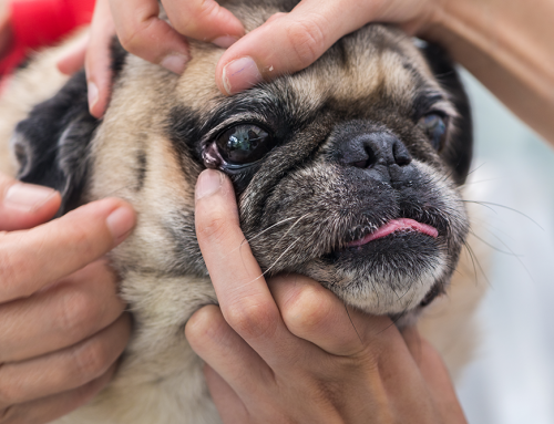
Lumps and bumps are one of the most common reasons families schedule veterinary appointments, and the concern behind that phone call is almost always the same: is this cancer? The honest answer is that no one can tell just by looking or feeling. A soft, moveable lump under the skin might be a harmless lipoma (a benign fatty mass), or it might be something that needs treatment. A firm growth on the gum might be an epulis (a benign oral mass) or a more aggressive tumor. The only way to know for certain is diagnostic testing, and the two most important tools we use for that are cytology (examining cells under a microscope) and biopsy (sending a tissue sample to a pathologist for a detailed analysis).
At Cobb & Co. Veterinary Clinic, we make cancer screening and early detection a priority. In addition to fine needle aspirates and biopsies, we offer Idexx CancerDx screening, a blood-based test that can help detect certain cancers before a visible mass even appears. Our surgical capabilities allow us to move from diagnosis to treatment efficiently when needed. If you have found a lump on your pet or simply want to stay ahead of potential problems, call us or book an appointment to get started.
Why You Can’t Tell by Looking
Signs of cancer in pets do not follow a predictable appearance. Malignant tumors can feel soft, smooth, and moveable just like a fatty lump. Benign masses can feel firm and tethered. Surface texture, size, location, and growth rate all provide clues but none of them provide a diagnosis. Cancer in pets affects one in four dogs over their lifetime, making diagnostic testing not an overreaction but a routine and important part of managing a lump discovered at any age.
Early testing means earlier answers. When something is caught at an early, smaller stage, treatment options are broader and outcomes are generally better.
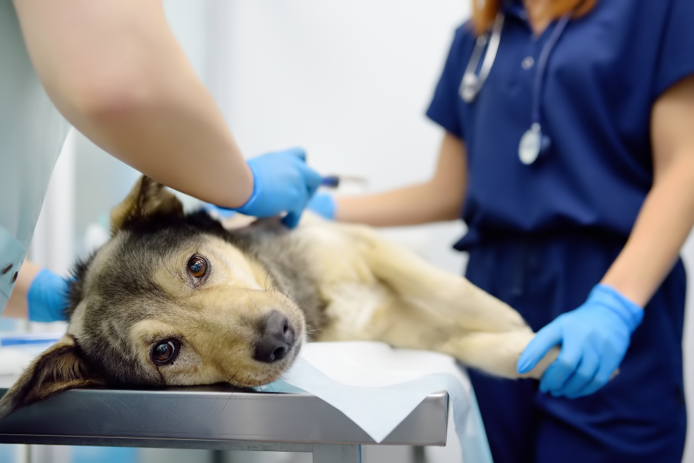
Fine Needle Aspiration: The First Diagnostic Step
How the Procedure Works
A fine needle aspiration (FNA) inserts a thin needle into a mass to collect cells or fluid, which are spread on a slide and examined microscopically. The process takes just a few minutes. Most pets tolerate it just like they do a vaccination. Multiple areas of a mass or multiple distinct masses can be sampled in the same visit.
Results from our reference laboratory typically return within a few business days. Our team explains findings in clear language and walks through next steps directly.
What FNA Can and Cannot Diagnose
Cytology from FNA examines individual cells rather than tissue structure. Skin cytology is especially informative for superficial masses and skin surface problems. It helps us differentiate between infection, inflammation, and normal vs. abnormal cells.
Some cancers, like mast cell tumors, are easily diagnosed with just an FNA. Other tumors release cells poorly into a needle. Fibrosarcomas, poorly exfoliating mammary tumors, and certain sarcomas may produce non-diagnostic samples even from a malignant mass. A non-diagnostic result never fully rules out cancer.
Skin Masses That Look Similar but Behave Very Differently
One reason cytology matters so much is that several common skin masses can look nearly identical on the outside but have completely different prognoses. Telling them apart by appearance alone is not reliable, even for experienced veterinarians.
Some of the most commonly confused masses in dogs include:
- Histiocytomas: benign, fast-growing, button-like masses that typically appear in young dogs. Many resolve on their own, but they often look alarming enough that families assume the worst. Cytology confirms the diagnosis in minutes.
- Melanocytic tumors: range from entirely benign pigmented growths to aggressive malignant melanomas, depending on location and cell characteristics. Oral melanomas are particularly aggressive and need fast, thorough evaluation.
- Cysts: benign fluid-filled or keratin-filled sacs that can feel firm, soft, or in-between. Sebaceous and follicular cysts are common and usually harmless, but some “cysts” turn out to be something else once cytology is done.
- Mast cell tumors: the most common skin cancer in dogs, and one of the best reasons never to skip testing a lump. They can look like just about anything, from a soft fatty-feeling bump to a red, angry-looking swelling, and they can change size from day to day. Cytology usually makes the diagnosis quickly, and grade and stage guide treatment from there.
- Lipomas: benign fatty masses that are extremely common in middle-aged and older dogs. They typically feel soft, moveable, and sit just under the skin. The catch is that they can feel identical to certain malignant tumors on physical exam, which is why even a classic-feeling lipoma is worth aspirating at least once.
Some of the most commonly confused masses in cats include:
- Feline basal cell carcinoma: one of the most common skin tumors in cats, often appearing as a raised, firm, or ulcerated mass. Typically slow-growing and often curable with complete surgical removal, but needs diagnosis first to distinguish from more aggressive tumors.
- Squamous cell carcinoma: often starts as a small scab or crusty area that keeps coming back, progresses to ulceration, and can become locally invasive if not caught early. These are especially important to evaluate in white or light-colored cats. Any skin lesion that has not healed within a few weeks warrants evaluation, not a wait-and-see approach.
- Fibrosarcomas: firm, locally aggressive tumors that most often develop in the skin and underlying tissue, sometimes at vaccine or injection sites. They tend to feel deep-seated and fixed rather than soft and moveable, and they are notorious for coming back if not removed with wide surgical margins. Biopsy is typically needed because these tumors often do not shed cells well on FNA.
- Mast cell tumors: behave differently in cats than in dogs. They often appear as raised, hairless, pink or reddish bumps on the head, neck, or trunk, and many of the cutaneous forms in cats are benign or behave less aggressively than the canine version.
The practical point: “it looks like just a fatty lump” or “it’s just a scab” is never a substitute for testing, because several of the conditions above can look exactly like a fatty lump from the outside.
Biopsy: When Tissue Architecture Matters
Types of Biopsy and How They’re Done
A biopsy collects tissue rather than individual cells, allowing the pathologist to examine cellular organization, invasion patterns, and structural relationships. These are only visible in tissue sections, not in individual cells on a cytology slide.
Common biopsy approaches:
- Punch biopsy: a circular cutting tool removes a cylinder of tissue; used for well-defined superficial masses
- Incisional biopsy: a wedge of tissue is removed from a mass without removing it entirely; used when diagnosis is needed before planning surgery
- Excisional biopsy: the entire mass is removed and submitted; combines treatment and diagnosis when the mass is small and surgically accessible
- Needle core biopsy: a larger-gauge needle collects a tissue core that preserves architecture without open surgery
All biopsy procedures require sedation or anesthesia to ensure patient comfort and accurate sampling. Tumor diagnosis through histopathology provides information cytology cannot.
What Histopathology Reveals
Histopathology is the diagnostic gold standard for cancer. The veterinary pathologist evaluates:
- Tumor type: the specific cancer confirmed to species and grade
- Grade: how aggressive the cells look and how they behave histologically
- Invasion: whether the tumor is infiltrating surrounding structures
- Margins: whether surgically removed tissue has clear margins (no tumor at the cut edge) or incomplete margins that may require additional surgery
Types of cancer that look identical on FNA may behave completely differently and require different treatments. Histopathology resolves that ambiguity.
When Biopsy Is the Only Way to Get an Answer
Several clinical situations highlight exactly why biopsy is sometimes the only path to a reliable diagnosis. A few worth knowing about:
Splenic Masses: Benign Nodule or Hemangiosarcoma?
When imaging finds a mass on the spleen, families often hear a frightening term: hemangiosarcoma. This is a reasonable concern because hemangiosarcoma in dogs is aggressive and common in certain breeds, particularly Golden Retrievers and German Shepherds. But not every splenic mass is hemangiosarcoma. Roughly half of splenic masses turn out to be benign (such as a hematoma or nodular hyperplasia) and the other half malignant. The two types can look identical on ultrasound, and FNA of a splenic mass carries bleeding risk and often produces non-diagnostic results because these masses are full of blood.
Histopathology after splenectomy (surgical removal of the spleen) is typically the definitive answer. This is why the standard approach for most actively bleeding or large splenic masses is surgery first, with the full spleen sent to the pathologist. A benign result gives families relief and a cure. A malignant result guides decisions about further staging and treatment.
Chronic GI Disease in Cats: IBD or Lymphoma?
Cats with chronic vomiting, diarrhea, and weight loss commonly have one of two conditions: inflammatory bowel disease or feline lymphoma. These two conditions can produce nearly identical clinical signs, nearly identical bloodwork findings, and nearly identical ultrasound appearances of a thickened intestinal wall. They are also related, because untreated IBD can progress to lymphoma over time.
The treatments, however, are completely different. IBD is managed with diet changes, immunosuppressants, and sometimes antibiotics. Lymphoma requires chemotherapy. Guessing wrong means either a cat with cancer who is not receiving effective treatment, or a cat with IBD receiving chemotherapy they do not need.
Biopsy is the only way to distinguish them reliably. This usually involves either endoscopic biopsy (samples taken through a scope) or surgical full-thickness biopsy (samples of the complete intestinal wall). Histopathology, sometimes combined with specialized testing like PARR (a test that looks at immune cell clonality), confirms the diagnosis and guides the right treatment.
Idexx CancerDx: Detection Before a Mass Appears
Beyond cytology and biopsy, Cobb & Co. offers Idexx CancerDx screening, a liquid biopsy platform that analyzes a blood sample for cancer-associated signals. This test is designed to detect certain cancers at an early stage, potentially before a visible mass is present.
CancerDx is particularly relevant for breeds with elevated cancer risk and for senior pets entering the demographic where cancer becomes more common. This is particularly important for breeds like Golden Retrievers, who are overly prone to lymphoma; this test can detect lymphoma up to 8 months before symptoms appear. A positive signal warrants follow-up imaging and targeted diagnostics to identify the source; a negative result provides reassurance as part of an annual wellness workup.
For families who want proactive early detection beyond physical examination alone, this screening adds a meaningful layer to annual wellness and preventative care visits.
Choosing Between FNA and Biopsy
What Guides the Decision
FNA is typically the first choice because it is fast, low-cost, and requires no anesthesia. Biopsy follows when:
- FNA returns non-diagnostic or inconclusive for a suspicious mass
- The tumor type requires grading for treatment planning
- Margin assessment after removal is clinically important
- Clinical suspicion for malignancy is high despite a benign FNA result
- The mass location makes FNA yield poor
What the Appointment Looks Like
For FNA: Bring your pet in during a regular appointment. The process takes a few minutes. Most pets go home immediately and results return within a few business days.
For biopsy: Pre-anesthetic bloodwork screens for metabolic concerns. The procedure takes 15 to 60 minutes. Your pet goes home the same day in most cases. An e-collar protects the site. Suture removal at 10 to 14 days. Histopathology results take one to two weeks.
In both cases, our team explains results in plain language and outlines next steps clearly. We do not leave you to interpret a lab report on your own.
Even a Benign Result Means Ongoing Monitoring
Here is something that often surprises families: a benign cytology result does not mean you never have to think about that lump again. Masses can change over months to years. Even benign masses, especially lipomas, can grow so large that they affect how your pet moves. Occasionally, the character of a mass genuinely changes over time, and what was once benign behaves differently.
This is why we recommend a simple monitoring routine for any mass, even one with a benign diagnosis:
- Measure the mass. Use a ruler or calipers to note length, width, and approximate height. Photos with a ruler next to the mass are also helpful for tracking.
- Write it down. Keeping a running list with the date, location, and size of each mass gives you and our team real data to compare over time.
- Check every month or two. A quick feel-over during a grooming session or belly rub is usually enough.
- Call us if anything changes. New color, new firmness, rapid growth, sudden size increase, ulceration, bleeding, or pain all warrant a recheck.
- Consider periodic re-aspiration. For masses that have changed in size, texture, or appearance, a repeat FNA is an easy way to confirm that nothing new is going on. Even without changes, some masses are worth re-aspirating every year or two as part of wellness visits, particularly in senior pets.
Monitoring does not need to feel obsessive. It is just a matter of staying familiar with what is normal for your pet so that change gets noticed early.
Acting on What We Find
Benign results: Provide a monitoring plan. Some benign masses are best left in place with periodic rechecks; others benefit from eventual removal when they reach a size that causes mechanical problems or start to change.
Malignant results: Open the conversation about surgical excision with margin goals, additional staging diagnostics, referral to an oncologist, radiation, chemotherapy, or palliative care depending on the specific diagnosis, stage, and your family’s goals.
Inconclusive results: Guide the next step, whether repeat sampling, biopsy, or imaging to characterize the mass further.
Frequently Asked Questions
How much does FNA cost vs. biopsy?
FNA is significantly less expensive. Biopsy costs more because of anesthesia and histopathology fees, but provides substantially more clinical information. We discuss the trade-offs specific to your pet’s situation.
Can I just watch the lump and see if it changes?
For some small, likely benign masses, monitoring with specific size-check intervals is appropriate. For rapidly growing masses, any mass accompanied by clinical signs, any mass where the index of suspicion for malignancy is meaningful, or any lesion that has not healed normally, testing is the right decision.
My pet’s lump aspirated as benign last year. Does it need testing again?
Not necessarily, but it should still be measured and monitored. If the mass has grown, changed in texture or appearance, or if new masses have appeared nearby, a repeat aspirate or biopsy is worth doing. For older pets, periodic re-aspiration of long-standing masses is reasonable even without obvious changes.
My pet seems completely healthy. Do I still need to test a lump?
Yes. Many malignant tumors are found in otherwise healthy-seeming pets before any systemic signs develop. Earlier testing means earlier treatment when it matters most.
Finding Answers Sooner
At Cobb & Co. Veterinary Clinic, early detection is something we actively pursue rather than wait for. Between Idexx CancerDx screening, fine needle aspirates, and biopsy capabilities supported by our surgical services, we can take a lump from an uncertain finding to a clear answer and a defined next step.
Book an appointment or contact us to get a mass evaluated or to ask about cancer screening as part of your pet’s next wellness visit.


Leave A Comment