
Vomiting, Weight Loss, and Diarrhea in Pets: A Guide to Diagnosing Intestinal Problems With Ultrasound
If your dog has been vomiting every few days, or your cat has lost a noticeable amount of weight without an obvious reason, you already know how frustrating it can be to not have answers. GI problems are among the most common reasons pets visit the vet, and one of the trickier ones to diagnose, because vomiting, diarrhea, and weight loss can point to a dozen different conditions that look nearly identical from the outside. Sorting through them takes a systematic approach, not guesswork.
At Cobb & Co. Veterinary Clinic, we work through GI complaints methodically, starting with a thorough physical exam and baseline diagnostics, then moving to abdominal ultrasound when we need a clearer picture of what is happening inside. Dr. Gilbert performs our abdominal ultrasounds in-house, and when imaging points toward something that requires a surgical fix, our surgical capabilities mean we can often handle the next step without sending you to a referral center. If your pet has been dealing with persistent digestive issues or unexplained weight loss, book an appointment so we can get to the bottom of it.
Signs Your Pet May Have a GI Problem
GI issues can show up gradually or seemingly overnight. Some pets vomit dramatically and obviously; others just slow down, eat a little less, and lose weight over weeks before anyone realizes something is wrong. Either way, the symptoms are worth taking seriously.
Signs that something is going on in the digestive system include:
- Vomiting:Occasional versus frequent, food versus bile versus blood- frequency and character both matter
- Diarrhea:Watery, bloody, or persistent diarrhea lasting more than 48 hours warrants a call to us
- Weight loss:Gradual loss despite a normal or even increased appetite often points to malabsorption- the gut is not absorbing nutrients the way it should
- Decreased appetite:Reluctance to eat, skipping meals, or turning away food that was previously a favorite
- Abdominal pain or bloating:Your pet may hunch up, guard their belly, be reluctant to move, or react when you touch their abdomen
- Lethargy:Not acting like themselves, lower energy, less interest in activity or interaction
- Blood in stool or vomit:Always warrants same-day evaluation
Any of these signs in isolation might be minor. In combination, or when they persist for more than a day or two, they tell us to look more closely.
Step One: Initial Diagnostic Testing
Before ultrasound enters the picture, we start with a physical exam and a set of baseline tests. These give us critical context and help narrow down what we are dealing with before we move to imaging.
Bloodwork gives us a broad overview of how the body is functioning. A complete blood count checks for signs of infection, inflammation, or anemia. A chemistry panel evaluates the liver, kidneys, and pancreas. Abnormal values do not always name the problem outright, but they point us in the right direction and tell us which organs to look at more closely.
Urinalysis evaluates kidney function and helps rule out urinary tract involvement, particularly useful when systemic illness or increased thirst and urination are part of the picture.
Fecal testing screens for intestinal parasites that can cause chronic diarrhea and GI upset, including giardia, hookworms, and roundworms. Even pets that have been on regular preventatives can occasionally test positive, so we do not skip this step.
Abdominal radiographs (X-rays) give us a broad structural overview of the abdomen- organ size and position, gas patterns in the intestines, and any obvious foreign material. They are particularly useful for spotting a large obstruction, significant intestinal gas dilation, or a dense object a pet has swallowed. X-rays do not show soft tissue detail the way ultrasound does, but they are fast, accessible, and often the first imaging step before we move to more detailed evaluation.
When initial testing comes back abnormal, or when it comes back normal and the symptoms keep going anyway, abdominal ultrasound is typically the appropriate next move.
What Abdominal Ultrasound Actually Does
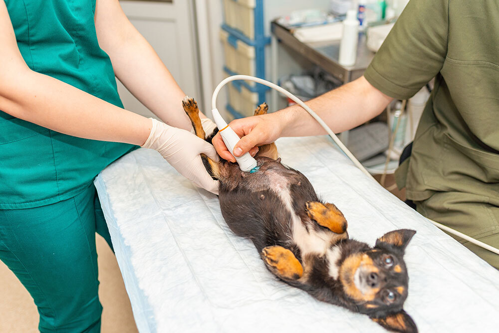
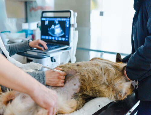
Ultrasound imaging works by emitting high-frequency sound waves that bounce off internal tissues at different rates depending on their density. A probe held against the skin captures those echoes and converts them into a real-time image on a screen. Unlike X-rays, which are excellent for evaluating bone structure and organ size but show limited soft tissue detail, ultrasound lets us see the texture, layering, and architecture of organs like the intestines, liver, stomach, and pancreas.
For most pets, the procedure involves shaving a small area of fur so the probe makes direct skin contact, then applying gel to eliminate air gaps between the probe and skin. It is non-invasive, uses no radiation, and the vast majority of dogs and cats tolerate it without sedation. Mildly anxious or painful patients may need light sedation to get a complete, accurate exam, but that is the exception rather than the norm.
Dr. Gilbert performs abdominal ultrasounds in-house at Cobb & Co., which means faster answers without an additional referral appointment.
When We Recommend Moving to Ultrasound
Ultrasound fills the diagnostic gap between “the bloodwork shows something off” and “we know exactly what is happening.” We typically recommend it when:
- Chronic vomiting or diarrhea has not resolved with initial treatment
- Bloodwork suggests organ involvement but does not identify the specific cause
- A mass or thickened tissue has been felt during a physical exam and needs characterization
- Unexplained weight loss continues despite dietary changes
- Abdominal pain or swelling is present without an obvious explanation
- A pet deteriorates rapidly and internal bleeding must be ruled out
If your pet has been showing any of these signs, a sick pet visit is the right starting point. We will examine your pet, run appropriate initial tests, and determine whether imaging is the right next step.
What Ultrasound Reveals: Common Intestinal Diseases and GI Conditions
Gastrointestinal Foreign Bodies and Blockages
Dogs in particular can be remarkably creative about what they decide to swallow, and when something cannot pass on its own, it becomes a problem quickly. Gastrointestinal foreign bodies cause obstruction when swallowed objects get stuck somewhere along the digestive tract. Ultrasound identifies the location of the blockage, the degree of intestinal dilation behind it, and whether surgical intervention is immediately necessary. This is one situation where having in-house imaging and in-house surgical capabilities at Cobb & Co. makes a meaningful difference in how quickly a pet gets the care they need.
Inflammatory Bowel Disease (IBD)
Inflammatory bowel disease is one of the most common causes of chronic vomiting and diarrhea in cats and dogs. It develops when the intestinal lining becomes persistently inflamed, disrupting normal digestion and the absorption of nutrients. On ultrasound, IBD often appears as thickening of the intestinal walls with disruption to the normal layered pattern. It can look similar enough to intestinal lymphoma that biopsy is ultimately needed to tell them apart, but ultrasound significantly narrows the diagnostic picture before any tissue sampling takes place.
Intestinal Lymphoma
Lymphoma affecting the GI tract often appears on ultrasound as diffuse intestinal wall thickening, enlarged abdominal lymph nodes, or distinct masses. It’s especially common in cats. Ultrasound identifies how much tissue is involved and where, and guides needle placement for cytology samples, providing important staging information before any treatment decisions are made.
Hemorrhagic Gastroenteritis (HGE)
Hemorrhagic gastroenteritis– now more commonly called acute hemorrhagic diarrhea syndrome, or AHDS- causes sudden, severe bloody diarrhea in dogs and can deteriorate into a dangerous situation without prompt treatment. Ultrasound helps rule out other causes of acute hemorrhagic GI signs, such as foreign body obstruction or intussusception (when one segment of intestine telescopes inside another), that require a different approach entirely. Distinguishing HGE from a surgical emergency early on matters enormously for outcome. If your dog is showing these symptoms, reach out to us right away; we are available for urgent care six days a week during open hours.
Pancreatitis
Pancreatitis– inflammation of the pancreas- is notoriously difficult to detect on physical exam alone, since the pancreas sits tucked behind other organs and cannot be easily felt. On ultrasound, an inflamed pancreas may appear enlarged with changes in echogenicity (how bright or dark the tissue looks on the image) and surrounding fat stranding, a sign of nearby inflammation. Abdominal pain, vomiting, and loss of appetite following a fatty meal are classic presenting signs, though pancreatitis does not always follow that predictable pattern.
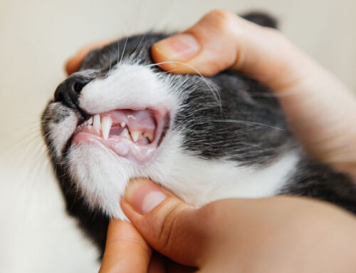
Hairballs and Chronic Vomiting in Cats
Cats who vomit frequently are not always “just being cats,” even if it can feel that way. While occasional hairball regurgitation is normal, frequent vomiting warrants a closer look to rule out IBD, lymphoma, hyperthyroidism, or other underlying GI conditions. Ultrasound can help distinguish between true structural GI pathology and recurrent hairball-related symptoms, which guides whether further diagnostic testing or simpler dietary and grooming changes are the better path forward.
Ultrasound-Guided Sampling: Turning Images Into Answers
One of ultrasound’s most practical capabilities is its ability to guide needle placement for tissue and fluid sampling. Rather than inserting a needle blindly toward an organ or mass, the probe visualizes the needle tip in real time, directing it precisely to the target.
Common sampling procedures guided by ultrasound include:
- Fine-needle aspirates (FNA):Rapid cellular sampling from a mass or organ to identify inflammation, infection, or cancer- often the step that definitively distinguishes IBD from intestinal lymphoma
- Cystocentesis:Sterile bladder taps that yield uncontaminated urine samples for accurate culture and urinalysis
- Abdominocentesis:Safe removal of abnormal abdominal fluid for analysis
Combining imaging and sampling in one visit reduces the number of steps between symptoms and clear answers, and our in-house capabilities mean we can process cytology quickly without unnecessary delays.
Putting It All Together: The GI Diagnostic Workup
No single test tells the whole story. The typical workup for a pet with chronic GI symptoms follows a logical sequence:
- Physical exam and history:Identify the pattern, timing, and severity of symptoms
- Bloodwork and urinalysis:Screen for organ disease, metabolic causes, and systemic illness
- Fecal testing:Rule out parasites as a contributing cause
- Abdominal radiographs:Evaluate organ size and position, intestinal gas patterns, and obvious foreign material
- Abdominal ultrasound:Assess GI organ structure in detail, identify masses, and guide sampling
- Tissue sampling if indicated:FNA or biopsy when imaging shows a suspicious lesion
- Diet trial if indicated:A controlled food elimination trial may be recommended when food sensitivity or allergy is suspected as a cause or contributing factor alongside structural GI disease
- Treatment and monitoring:A plan tailored to the confirmed diagnosis, with follow-up imaging as needed
Our approach at Cobb & Co. is to work through this process with you, explaining our reasoning at each step so you understand what we are looking for and why.
Supporting GI Health During and After Diagnosis
For pets managing GI conditions identified through diagnostics, nutritional and probiotic support are often part of the long-term plan. FortiFlora and Proviable are widely used veterinary probiotics for dogs and cats that support intestinal microbiome balance during GI recovery.
For cats with hairball-related vomiting, Laxatone and hairball control soft chews can help reduce frequency, and hairball care diets are also available.
Frequently Asked Questions About Veterinary Ultrasound and GI Issues
Does my pet need to be sedated for an abdominal ultrasound?
Most pets do not require sedation. Light sedation may be used for anxious, painful, or fractious animals to ensure a thorough and accurate exam, but it is not routinely necessary.
Is abdominal ultrasound safe?
Yes. Ultrasound uses sound waves, not radiation, and has no known harmful effects with standard diagnostic use.
Can ultrasound diagnose cancer definitively?
Ultrasound identifies masses, lymph node changes, and organ abnormalities consistent with cancer, but a definitive diagnosis always requires tissue sampling. Ultrasound guides where to sample and provides important staging information, making it a critical part of the process even when biopsy is the final step.
How is ultrasound different from an X-ray?
X-rays are well-suited for evaluating bone, chest structure, and organ size. Ultrasound provides real-time images of soft tissue architecture and internal organ structure in much greater detail. The two tools are complementary, and we often use both as part of the same workup.
How do I know if my pet needs an ultrasound?
If your pet has chronic vomiting or diarrhea, unexplained weight loss, abdominal pain or swelling, or bloodwork pointing to GI or organ disease, ultrasound is often the appropriate next step. A conversation during a sick pet visit will clarify whether imaging makes sense for your pet’s specific situation.
Getting to the Bottom of Your Pet’s Intestinal Issues
GI problems are some of the most common and most diagnostically challenging conditions we see, precisely because so many different diseases produce the same outward symptoms. Abdominal ultrasound gives us the ability to look inside your pet without surgery and without radiation, converting vague and persistent symptoms into specific, actionable findings.
At Cobb & Co. Veterinary Clinic, we perform abdominal ultrasounds in-house, and our wellness and preventative care approach means we are invested in catching problems early and managing them well over the long term. If your pet has GI symptoms that have not been fully explained, contact us or book an appointment and let’s figure out what is actually going on.


Leave A Comment